Candidates in context
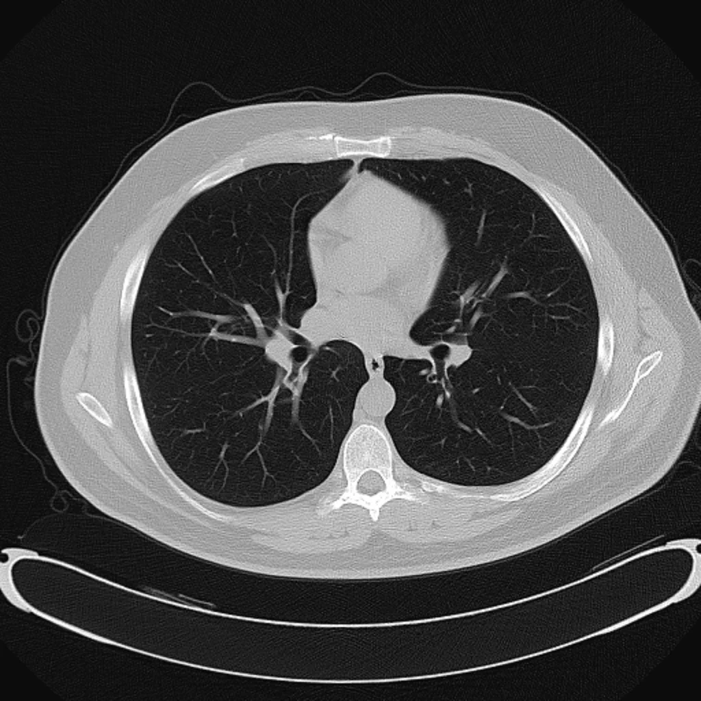
Move from a suggestion to its location in the CT study.
AI-ASSISTED LUNG CT ANALYSIS
Pulmonary nodule intelligence, built around the radiologist. Bringing potential findings into focus, with a thoughtful AI-assisted review workflow.
THE CLINICAL CONTEXT
Chest CT brings extraordinary detail—and a demanding volume of information. Radiologists balance careful visual review with the pace and complexity of everyday practice.
We’re building an additional layer of attention, so expertise can stay focused on interpretation.
A complementary layer of attention.A THOUGHTFUL SECOND READER
Specialized AI, working together. Clinical judgment, always with you.
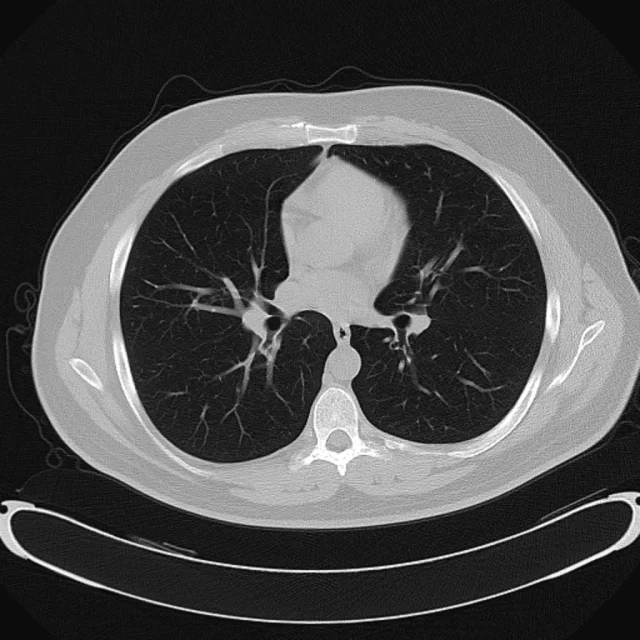
A detection agent is being developed to examine chest CT images and identify potential pulmonary nodule candidates.
Potential findings brought into viewCOLLABORATIVE INTELLIGENCE
Specialized AI agents, each with a clear role. A detection layer looks broadly. A verification layer considers the candidates. You bring the clinical perspective.
Our approach to intelligenceTHE PLATFORM EXPERIENCE
Explore an illustrative review experience. Move between candidates, inspect their location and make your own assessment—directly in the study.
Try selecting a candidate below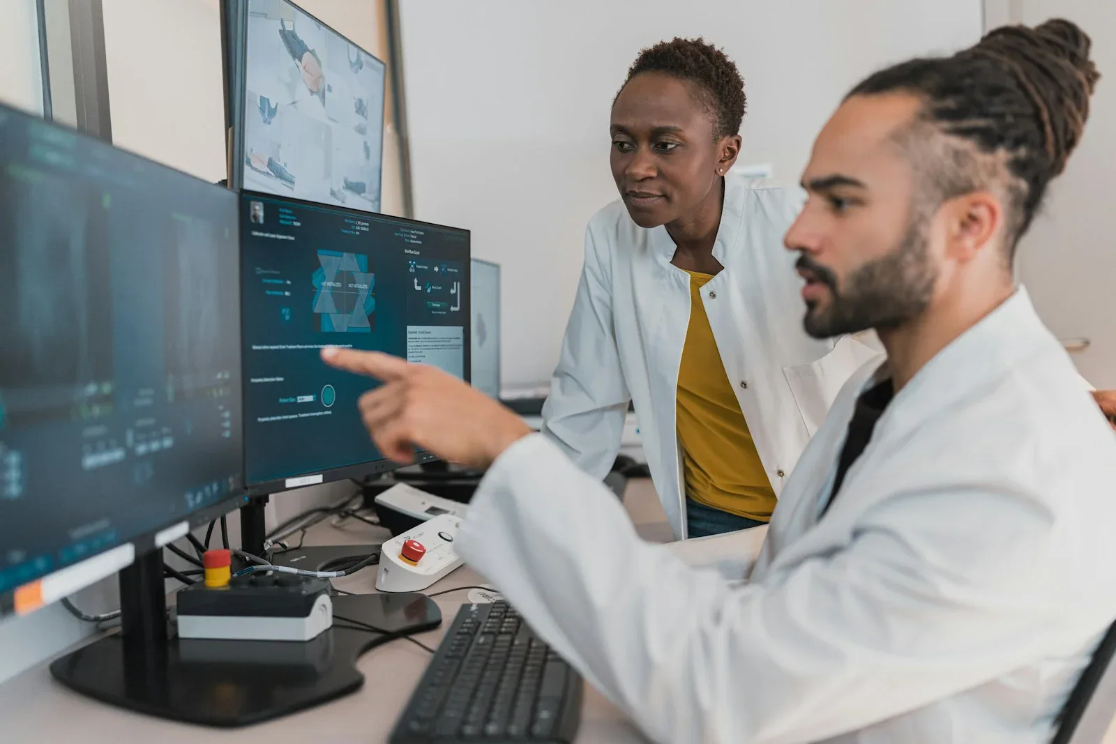
DESIGNED FOR RADIOLOGISTS
A useful assistant should fit the way you think, see and review. That is where our design begins.
Move from a suggestion to its location in the CT study.
A considered candidate list, designed to support attention.
Inspect, accept or dismiss. The interpretation is yours.
Visible regions and clear review states, rather than a hidden verdict.
OUR GUIDING PRINCIPLE
Medical imaging is more than pattern recognition. It is experience, context and clinical judgment. We’re building AI to extend that expertise, with every suggestion open to review and every decision kept human.
EVIDENCE BEFORE ASSURANCE
Trust begins with a clear understanding of what a system can—and cannot—do. Our evaluation framework is being shaped around that responsibility.
Pending validation
Pending validation
Pending validation
Pending validation
A FOCUSED START. A BROADER VISION.
We’re building a foundation for imaging intelligence, one carefully evaluated capability at a time.
A focused starting point for AI-assisted chest CT review.
01 / FOUNDATIONA specialized layer to assess potential findings in context.
02 / REFINEMENTExploring comparison over time and future clinical decision support.
03 / HORIZONPlanned capabilities are subject to research, validation and applicable regulatory requirements.
FOR HOSPITALS & IMAGING CENTERS
Useful intelligence must also work in practice. We’re considering scalable analysis, deployment flexibility and future PACS/RIS interoperability from the beginning.
Explore clinical collaborationA design direction for future clinical interoperability.
An architecture intended to support growing study volumes.
AI findings remain visible and open to clinician assessment.
THE COMPANY WE’RE BUILDING
Pulmonary nodule detection is our starting point. Our ambition is a broader platform that helps clinicians find meaningful information in increasingly complex imaging data.
Meet our missionMEET OUR TEAM
Artificial intelligence, biomedical engineering and clinical radiology. Different perspectives, united by a clearer vision for medical imaging.

Senior Computer Vision Engineer
Senior Biomedical Engineer
Senior Biomedical Engineer
Radiologist
LET’S BUILD THE NEXT CHAPTER
We’re looking to collaborate with radiologists, imaging centers
and clinical research partners.